The patient is a 58-year-old diabetic male who in August of 2022 suffered a penetrating injury of a rusty nail to the plantar calcaneus of the right foot, subsequently developing a subacute osteomyelitis. The wound was closed successfully; however, over the course of the next several months, the patient developed an infected bone cyst of the plantar medial calcaneal tuber which resulted in a pathologic fracture of the calcaneus. At that time, the patient’s hemoglobin A1c was approximately 12%, and he was immobilized in a below-knee cast as the patient’s blood glucose became well controlled. Serial imaging was performed in the form of an x-ray, MRI and CT scan. Most recent CT scan demonstrated worsening of the pathologic fracture and cystic lesion at the plantar medial tubercle of the calcaneus. As such, with the patient’s hemoglobin A1c now reduced to approximately 6%, the patient elected to undergo corrective surgery for this condition in the form of debridement of all infected material from the pathologic fracture and bone cyst, insertion of antibiotic, ceramic and application of lower extremity external fixator for stabilization and to prevent further displacement of the pathologic fracture.
The cystic lesion was approached from a posterior angulation immediately inferior to the distal fibers of the Achilles tendon and an angulation of approximately 30 degrees. Ultrasonic diamond tip bur was used to penetrate the cortex of the posterior calcaneus and drained the cyst which was cultured and sampled for pathology. The cystic lesion was then loaded with antibiotic impregnated bone ceramic (calcium phosphate). Following successful completion of this portion of the procedure the lower extremity external fixator was then applied in order to stabilize the extremity and prevent pathologic fracture.
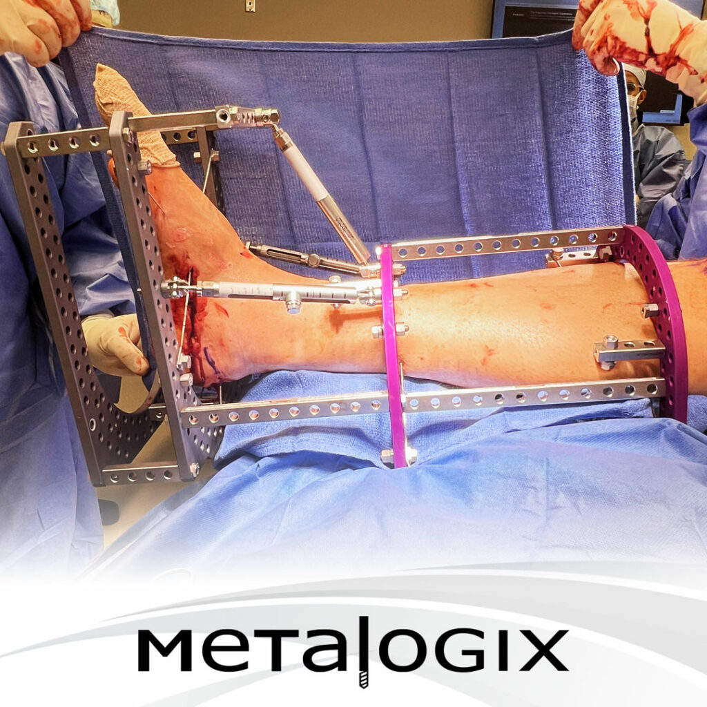
The base construct of the external fixator involved tibial plates connected by spanning posts within all wire techniques performed for maximum stability and prevention of stress risers. The tibial block was then connected to the footplate using a combination of posts and super struts. The footplate was placed superior to the cyst in order to avoid wire placement directly through the cyst and as such a secondary footplate was added for increased stability below the construct. Additionally the footplate was closed off with posts and connected to the tibial block using a super struts this is a completely static construct with the purpose of maximum stability. The intention is for the fixator to be left on for approximately 12 weeks, at the 6-week mark a repeat CT scan will be taken as well as blood inflammatory profile in order to determine if the antibiotic laden bone ceramic can be removed and replaced with autograft bone graft. The procedure was tolerated well and made successful by the revolution system allowing for maximum stabilization with open posterior back to accommodate for edema in the post procedure period.
The procedure was tolerated well by the patient with the expectation of stageII occurring in approximately 6 weeks


